

")
Removal of moles
Almost every adult has a number of moles
The common mole is completely harmless.
The nevus is a common benign skin lesion due to local proliferation of pigment cells (melanocytes). It is more properly called a melanocyte nevus. A brown or black spot contains melanin.
A nevus may be present at birth (congenital nevus) or appear later (acquired nevus).
Almost everyone has at least one mole
About 1% of people are born with one or more congenital melanocyte nevi. This is usually sporadic.
People with light skin tend to have more nevi than darker ones.
Neonatal nevi (2 to 10 years old) tend to be the most obvious and persistent throughout life.
Males acquired later in childhood or adulthood often follow sun exposure.
Diagnosis
Nevi are usually diagnosed clinically by their typical appearance. An experienced dermatologist alone is the one who diagnoses and maps them. We must be very careful if:
A mole changes size, shape, structure or color
A new nevus develops in adulthood (& gt; 40 years old)
It looks different from other moles (the so-called ugly duckling)
It has ABCD features (Asymmetry, Border Irregularity, Color Variation, Diameter & gt; 6 mm)
There is bleeding, crusting or itching.
Most skin lesions with these characteristics are actually harmless when evaluated by a dermatologist. A detailed nevus mapping is the best solution for high risk individuals
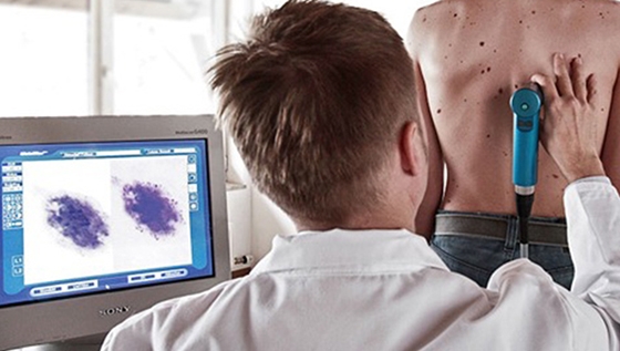
Mole Mapping
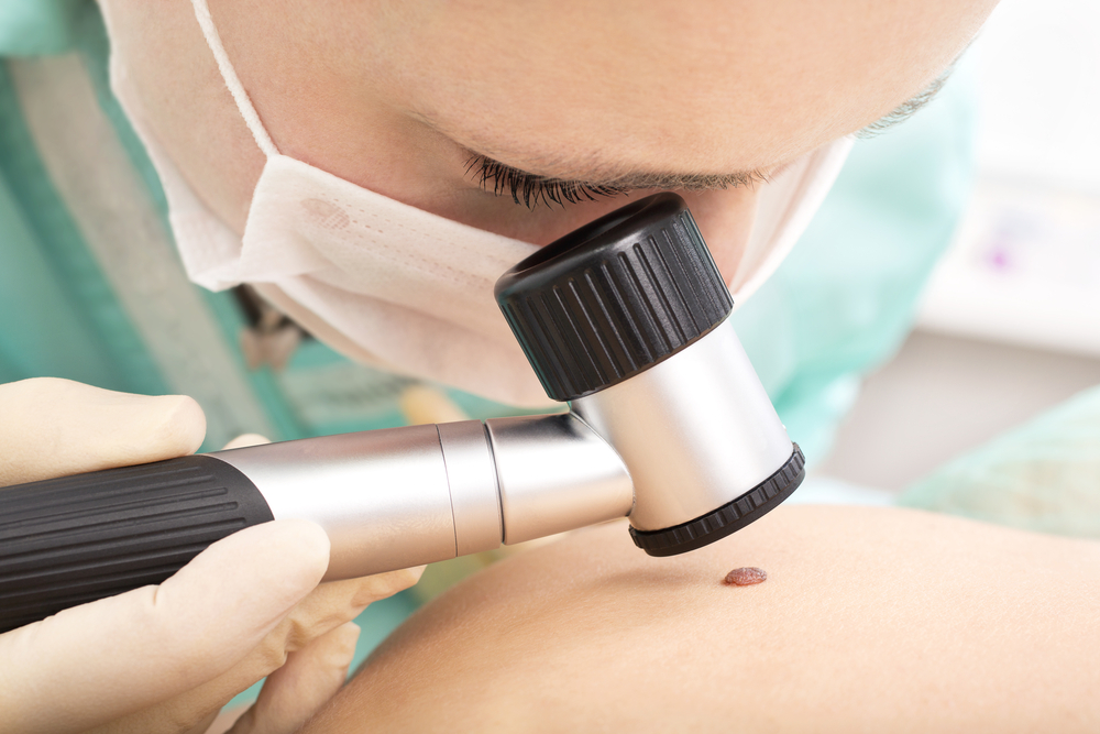
Dermatoscopy is a painless and harmless method by which we mainly examine brown lesions. We use this tiny magnification process to find out if a pigmented area of skin is benign or suspicious.
In digital dermatoscopy we do not work with a hand-held optical microscope, but use special digital images. We use this method when we do not intend to remove a colored lesion immediately, but we want to observe its further course exactly.
Colored lesions that do not change over time are less suspect. Digital dermatoscopy makes the slightest changes visible under standard conditions.
Since suspicious moles are often surgically removed and then proven to be benign lesions, this method can save us from unnecessary interventions.
Skin cancer
"Skin cancer" summarizes a variety of different types of skin cancer. The most dangerous is black skin cancer, malignant melanoma. Melanoma affects all ages and is responsible for most deaths among all cancers. His white cancers are much more common 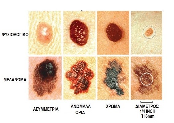 skin, basal cell carcinoma, squamous cell carcinoma and its precursor stages, and occur mainly in the elderly. In addition, there are other very rare forms of skin cancer. Other malignant changes in the skin may represent skin metastases of cancer of the internal organs. Thus, a change in the skin can lead to the diagnosis of a hitherto unknown metastatic cancer.
Basal cell carcinoma - Squamous cell carcinoma
Basal cell and squamous cell carcinomas can be summarized as white skin cancers. Both types are derived from cells of the upper layer of the skin, the epidermis. Basal cell carcinoma is a common disease in older men. But both women and young people can show it. Appears on the bridge of the nose and the rest of the face. A subtype, superficial basal cell carcinoma often occurs in the trunk. Basal cell carcinoma develops slowly, destroying healthy tissues but not metastasizing to other organs. . It is a glowing nodule in the color of the skin,with varicose veins and is surrounded by very small pearly nodules. Sometimes in the center it creates an ulcer that bleeds. The treatment is surgical removal.
Squamous cell carcinoma is also a disease of the elderly after many years of sun exposure, and occurs in particularly sun-exposed areas of the face: ears, lower eyelids, lower lip, scalp in the elderly who have lost their hair.Its precancerous lesions are called radial hyperkeratosis. They are flat, reddish lesions that peel off and cause mild itching. During the transition to carcinoma, they gain weight, become nodular, and bleed. The treatment is surgical removal. Unlike basal cell carcinoma, it can (extremely rarely) metastasize to distant organs through the lymphatic system. However, if detected early it is completely cured by removal.
Malignant melanoma
Malignant melanoma (black skin cancer) originates from skin pigment cells. As a result, the color is usually black, but there are also light-colored variants (melanoma melanoma), which are particularly difficult to detect. Most melanoma develops in the trunk and limbs, sometimes from pre-existing moles, sometimes from new ones. They can also appear on the face (especially in the elderly), on the nails, soles, mucous membranes, eyes. Men and women are equally affected, often between the ages of 50 and 60. However, melanoma also occurs in young people aged 16 and over. The risk increases with tumor thickness. This is measured in the histological preparation. In thin melanomas with a tumor thickness of less than 1 mm, the risk of metastasis is low. In thick melanoma over 4 mm on the other, there is a high risk of metastasis. Today, most melanoma is fortunately diagnosed in time and thus removed with a small tumor thickness. About 85% of patients with melanoma are treated with the first removal.
Who should have a regular nevus test
Number of nevi over 50
Family history of melanoma
Individual history of skin cancer or melanoma
Light skin phototype (phototype 1-2)
Individual history of sunburn in childhood or adolescence
People with giant congenital melanocyte nevus
Dysplastic nevus syndrome.